WellVu Thermal Cameras allow you to take a picture of your patient’s physiological health in real-time, what is happening right at that moment. All other imaging that we do in practice, radiographic, ultrasound, MRI, and CT studies, tell us what has already happened.
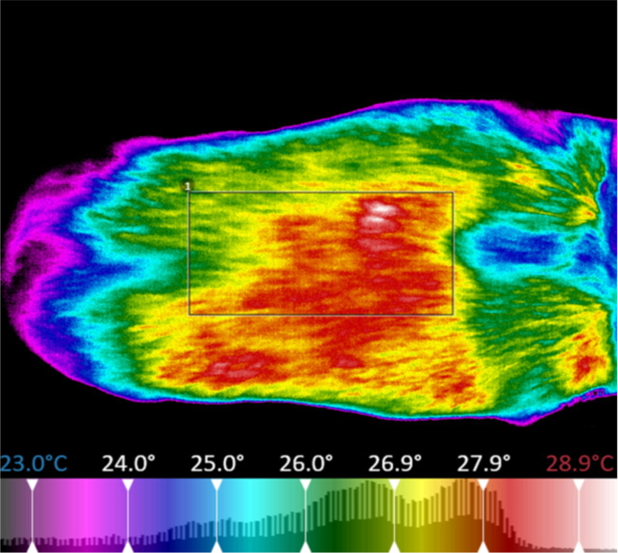
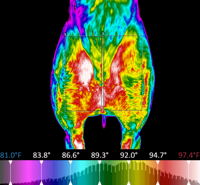
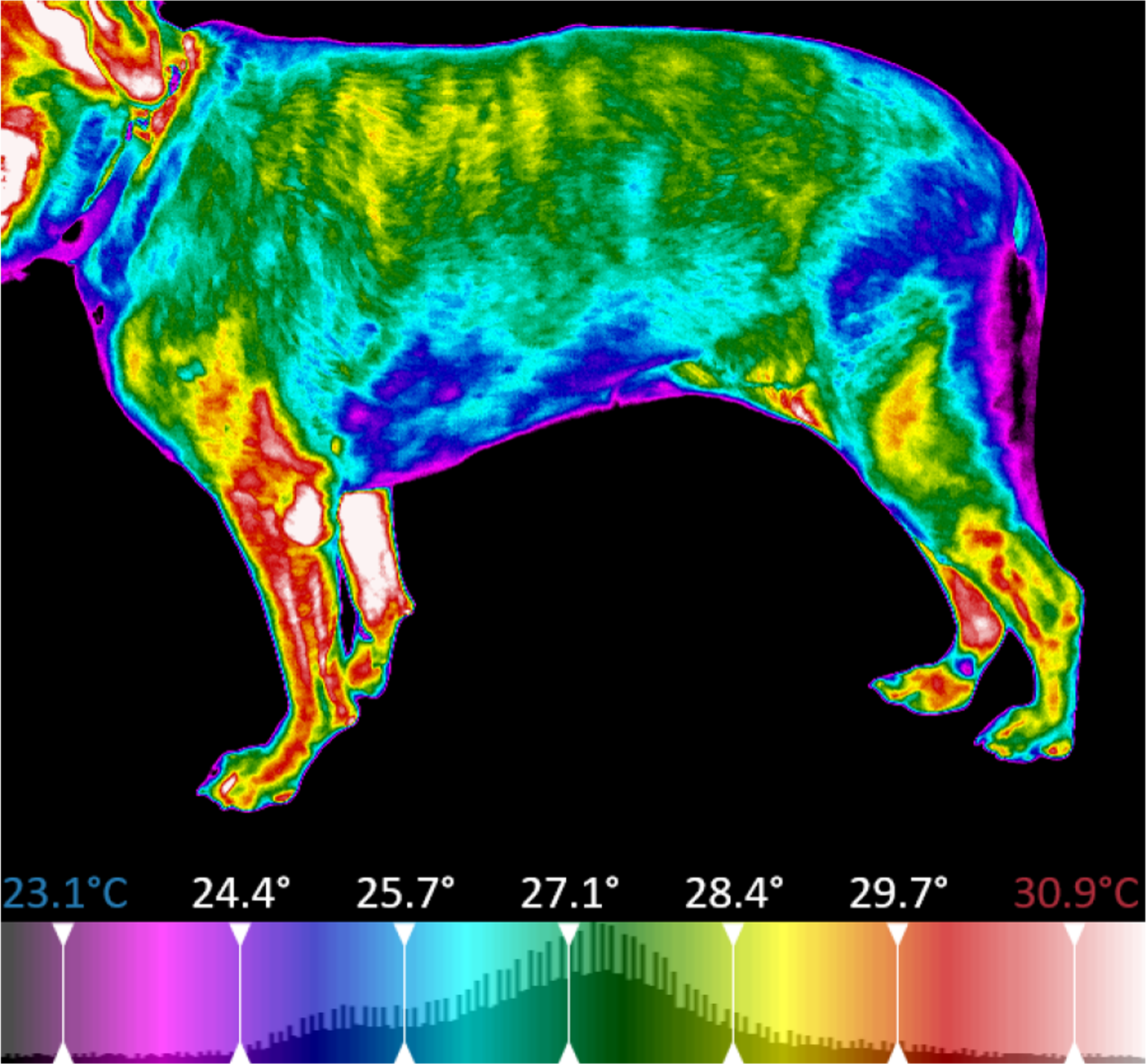
Thermal energy is constantly being emitted from our patients. The amount of energy is dependent on the anatomical location, the vascular supply to that area, and its neurological state.
Nature is symmetrical; look at a leaf or snowflake. When our patients were younger, they were more thermally symmetrical when considering contralateral anatomical areas. As time marches on, our patients become thermally asymmetrical due to injury, disease, and aging. Identifying these areas of thermal asymmetry is an indication that there is an issue that deserves further investigation and, potentially, diagnostics.
WellVu thermal imaging system's high resolution and sensitivity allows you to instantly visualize the physiological state of your patient. Medically calibrated veterinary-specific software compiles this radiated temperature data, converts it to an electrical impulse, and finally produces a detailed image: a thermogram. This thermogram is an easy-to-interpret illustration of the physiological state of the patient. The patient is its own control so any thermal asymmetry should warrant further investigation.